Bunion formation is one of the most common disorders of the foot, yet most people have an incomprehensive understanding of it. It is for this reason that bunion images have become so important for educating patients and helping podiatrists understand this condition for better diagnosis and treatment. This article will look at the significance of bunion images in taking a patient through identification to the recovery stages of bunions.
Imaging is a cornerstone in medical diagnostics, and so it is with bunions. Bunion pictures serve to allow patients an insight into the stages of this condition and compare that to their symptoms. In this respect, these pictures are very important for podiatrists in assessing the degree of a bunion and establishing appropriate treatment recommendations.
Whether it’s a clinical photograph, an X-ray, or a post-treatment visual, each type of image helps in understanding the bunion’s impact on foot anatomy. Additionally, they can demystify medical jargon, helping patients feel more confident about their treatment choices.
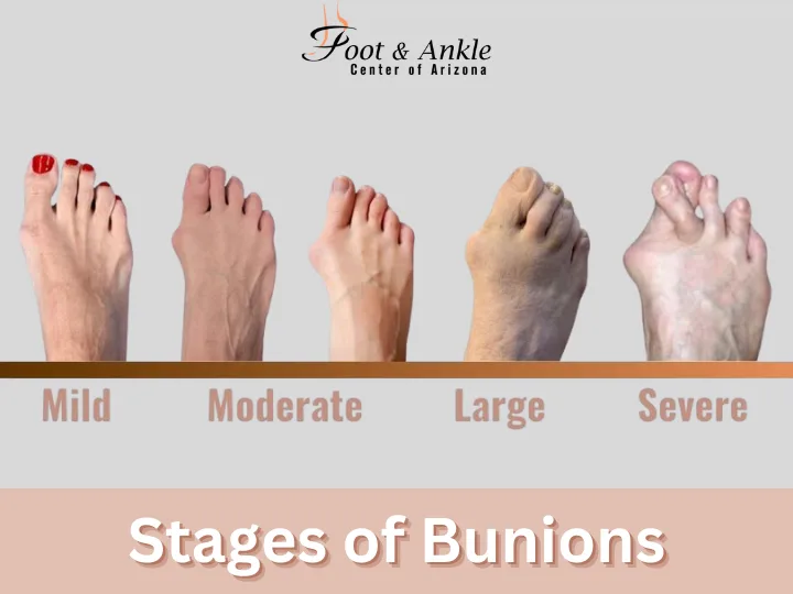
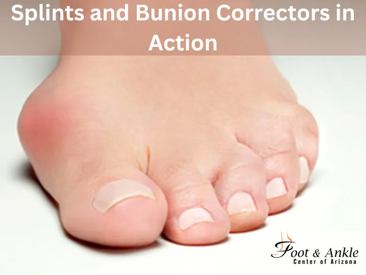
Conditions involving bunions range from minor inconveniences to severe deformities that impact daily life. Bunion images serve to diagnose these stages:
These images provide a clear comparison, helping the patient identify where they fall on the severity spectrum.
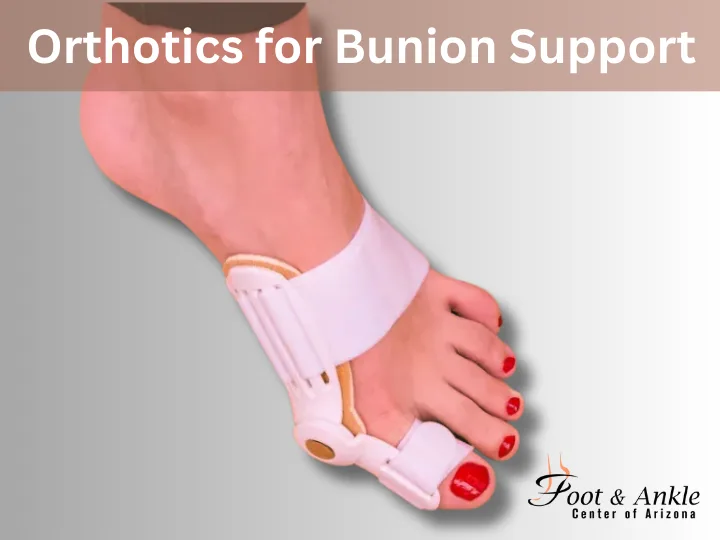
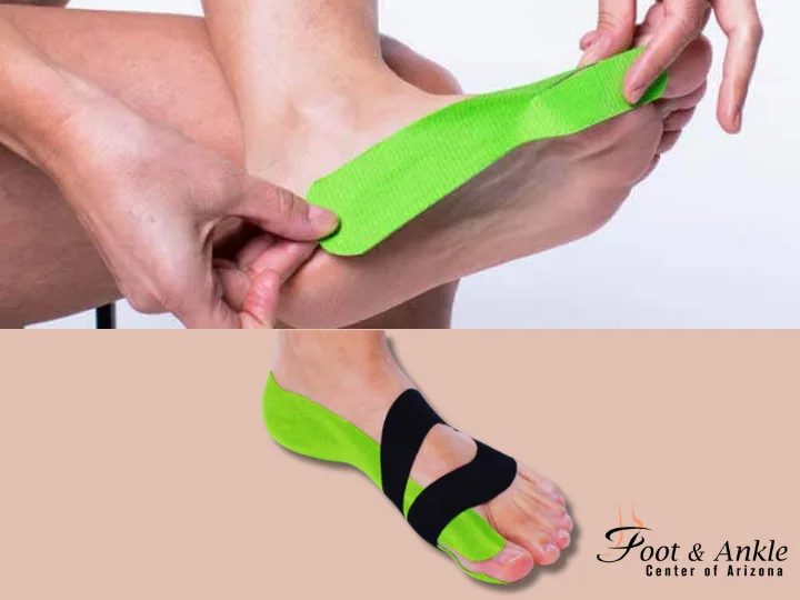
Non-surgical approaches are often the first line of defense in managing bunions. Bunion images can help illustrate the effectiveness of these treatments:
Images of proper footwear demonstrate how shoes with wider toe boxes and better arch support can alleviate pressure on the bunion. Comparisons with improper footwear underline the importance of making the right choices.
Custom orthotics offer visible changes in foot alignment. Side-by-side images show how these inserts redistribute weight and reduce strain on the bunion area.
Step-by-step visuals highlight how padding protects the bunion from irritation while taping techniques provide support to misaligned toes.
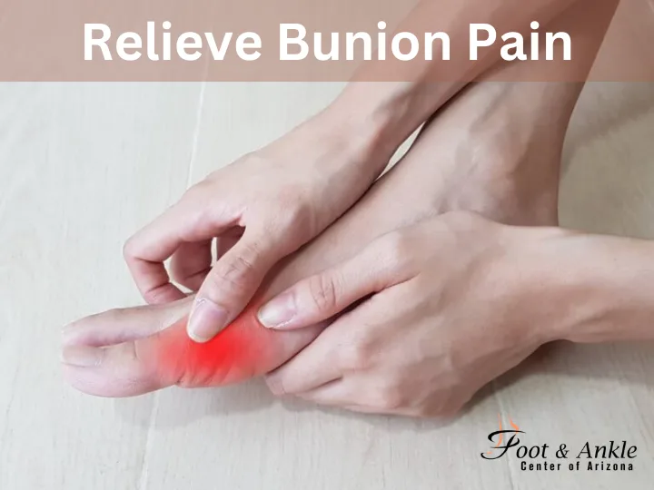
Images of foot relaxation exercises, such as toe stretches or massages, show patients practical ways to ease bunion pain.
Therapeutic exercises, illustrated with bunion images, help strengthen foot muscles and improve mobility.
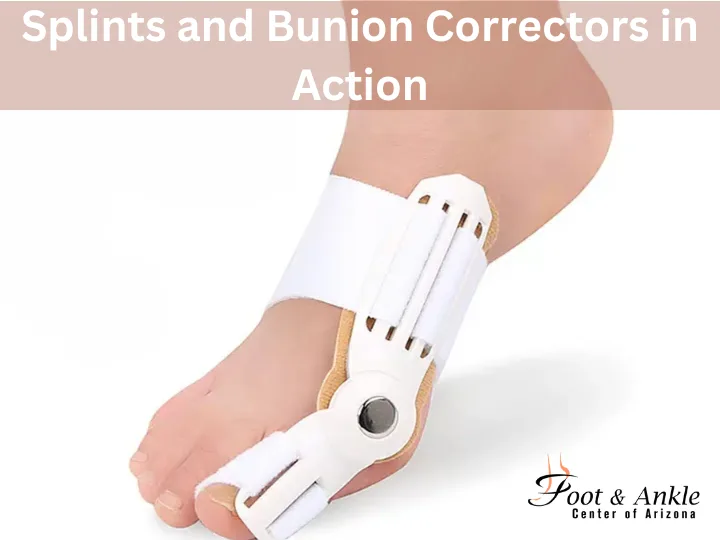
Photos of splints demonstrate how these devices realign the big toe, providing relief during sleep or daily activities.
When non-surgical methods fail, surgery may be necessary. Bunion images provide a clear understanding of various surgical options and their outcomes.
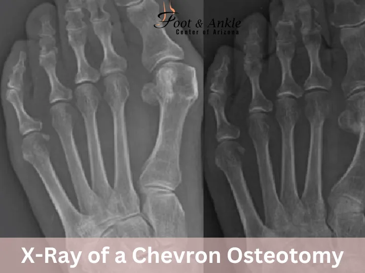
This procedure corrects mild to moderate bunions by realigning the metatarsal bone. Before-and-after X-rays show significant improvements in toe alignment.
Images demonstrate how this procedure reduces tension around the joint, restoring a more natural toe position.
Before-and-after images showcase the removal of excess bone growth, revealing a smoother foot profile.
Each type of osteotomy is visually distinct, with X-rays showing how bones are repositioned to correct alignment.
Images highlight the fusion of bones in severe cases, improving stability and reducing pain.
Visuals of the midfoot area before and after surgery illustrate the procedure’s effectiveness in correcting deformities.
Photos demonstrate how this procedure removes part of the affected joint, especially in cases of severe damage.
Small incisions and faster recovery are key benefits of MIS. Images show minimal scarring and significant post-surgery improvement.
Recovery is a critical phase in bunion treatment, and bunion images help track progress over time.
Images taken immediately after treatment, mid-recovery, and upon full healing show the patient’s journey. These visuals highlight improvements in foot shape and functionality.
Photographs document key milestones, such as reduced swelling, restored alignment, and regaining mobility. For patients, these images provide reassurance and motivation during the recovery process.
Imaging is critical for both diagnosis and treatment planning of bunions. High-quality bunion images, both X-rays, and clinical photographs, provide great details about the nature of the deformity. Patients find these images helpful, often utilizing them to document the changes in their condition over time and the results of treatments.
Imaging is also utilized by podiatrists to identify complications, such as joint damage or arthritis, which may require more specialized care.
Visuals are very important for patient education. Bunion images simplify the more complex medical concepts and allow patients to make informed choices.
For example, operation diagrams like Chevron or Lapidus techniques allow a patient to know what to expect both during and after the surgery. Pictures of before and after truly show the real outcomes, enabling confidence in treatment plans.
Moreover, exercise and taping techniques images provide patients with instructional steps that they can utilize at home to help them manage their condition.
Bunion images are much more than visual aids; instead, they are strong tools for diagnosis, treatment planning, and patient education. From assessing the severity of a bunion to monitoring recovery, images provide clarity and confidence for patients moving through the bunion journey.
If you’re struggling with bunion pain or seeking guidance on treatment options, consult a podiatrist who can provide expert advice and imaging. Visit our clinic or website today to explore comprehensive bunion care supported by the latest imaging technology.