The ankle is a complex joint integrating the lower end of the tibia (shinbone) and the superior aspect of the talus (anklebone). The talus has a dome shape at its top, which is thoroughly coated with cartilage, a durable, resilient tissue that facilitates smooth ankle motion. An injury to this cartilage and the underlying bone on the talus inside the ankle joint is known as a talar dome lesion. Other terms for this condition include osteochondral defect (OCD) and osteochondral lesion of the talus (OLT), with “osteo” referring to bone and “chondral” indicating cartilage.
These lesions typically result from an injury, like an ankle sprain. If the cartilage fails to recover adequately after the injury, it may soften and start to fragment. Occasionally, a piece of the deteriorated cartilage and bone breaks away and floats within the ankle joint.
The development of symptoms can be slow and may take various months or even years post-injury. Common symptoms include:
Diagnosing a talar dome lesion can be tricky as pinpointing the precise pain location is often difficult. The process involves the foot and ankle surgeon asking the patient about past injuries and performing an examination to check for pain, clicking, or restricted motion in the ankle.
Diagnostic measures may include an anesthetic injection into the joint to see if the pain subsides temporarily, indicative of joint-related pain. X-rays and possibly MRI or other imaging tests are typically ordered to assess the lesion and injury extent.
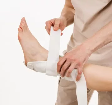
The approach to treating a talar dome lesion depends on its severity. For stable lesions, without loose cartilage or bone pieces, treatment might include:
Surgery may be the next step if the lesion doesn’t improve with nonsurgical treatments. The surgery aims to remove any bone and cartilage fragments and create a conducive healing environment. The optimal surgical technique is determined based on the individual case.
Arthritis might develop in the ankle joint due to cartilage damage, leading to chronic pain, swelling, and limited motion. Management of these issues is typically guided by a foot and ankle surgeon and may involve: